flow cytometry results for lymphoma
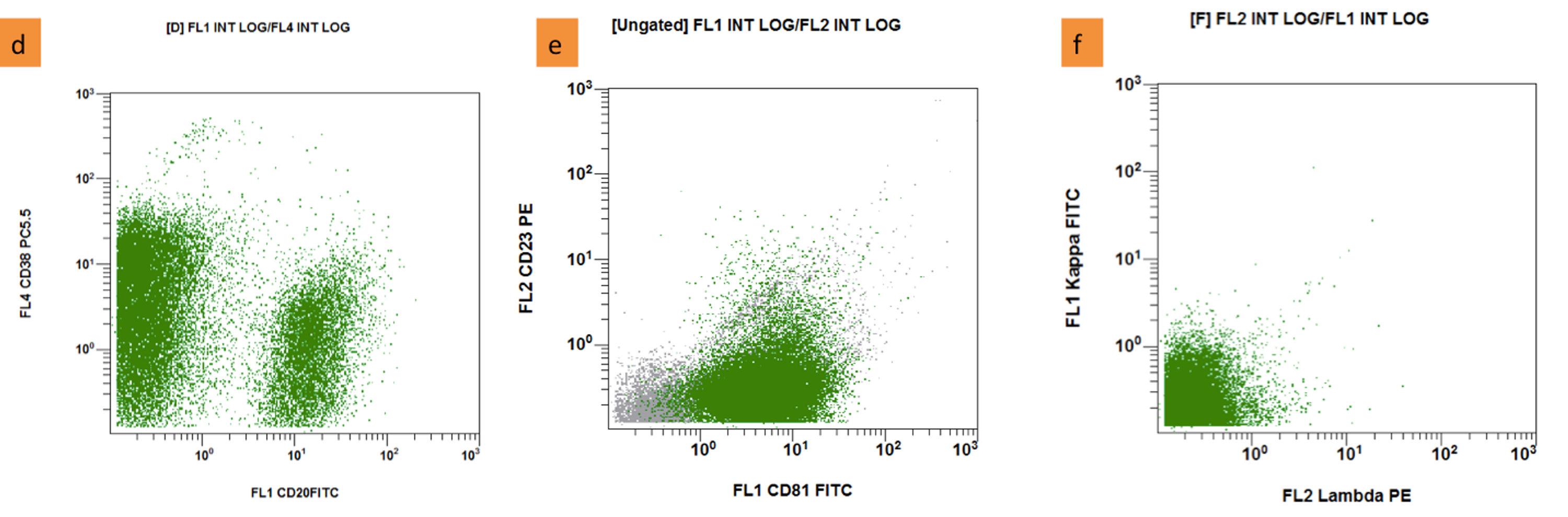
Flow cytometry is rapid and appears to be virtually diagnostic of non-Hodgkins lymphoma when a majority of cells are B cells with an abnormal kappalambda ratio. Quantification of minimal residual disease may guide therapeutic strategies in mantle cell lymphoma.

Flow Cytometric Immunophenotyping Performed On The Same Plasmablastic Download Scientific Diagram
Grade 1 follicular lymphomas had a percentage of cells at or beyond the 500-channel mark ranging from 012 to 66 median 46 whereas grade 2 follicular lymphomas had a percentage ranging from 412 to 1255 median 7.
. Expert Answers to Your Questions. Eight cases 32 were detected by flow cytometry alone and were missed by histomorphology analysis and 6 of these 8 cases showed minimal bone marrow involvement 009-22. 5 segs 52 lymphocytes 32 monocytes 9 eosinophils.
47XX8t922q34q112146XX19 t922 translocation in 1 of 200 cells analyzed. While multiparameter flow cytometry is used for diagnosis the gold standard method for minimal residual disease analysis is real-time quantitative polymerase. The added clinical value is the speed by which flow cytometry can establish or confirm the diagnosis enabling a faster initiation of treatment while false positive cases were.
The Symptoms Of Lymphoma. Flow cytometry has become an important tool in the diagnosis of mature lymphoid neoplasms and the determination of prognosis in selected cases. Flow cytometric leukemia and lymphoma analysis may aid in identifying the tumor lineage for diagnostic and prognostic purposes.
It is used to detect abnormal hematolymphoid populations determine what cell surface markers they express and integrate immunophenotypic findings with morphologic and available clinical and. Ad Discover the Latest Transplant Treatment Options at Mayo Clinic. Flow cytometry is generally used as follow up testing after a complete blood count CBC or white blood cells scan WBC.
Ad Discover These Tips To Help Relieve The Symptoms And Signs Of Lymphoma. Flow cytometry analysis in brain biopsy is a feasible technique with 100 specificity to confirm the diagnosis of brain lymphoma in patients suspected for lymphoma on clinical grounds. CSF samples from 105 patients with newly.
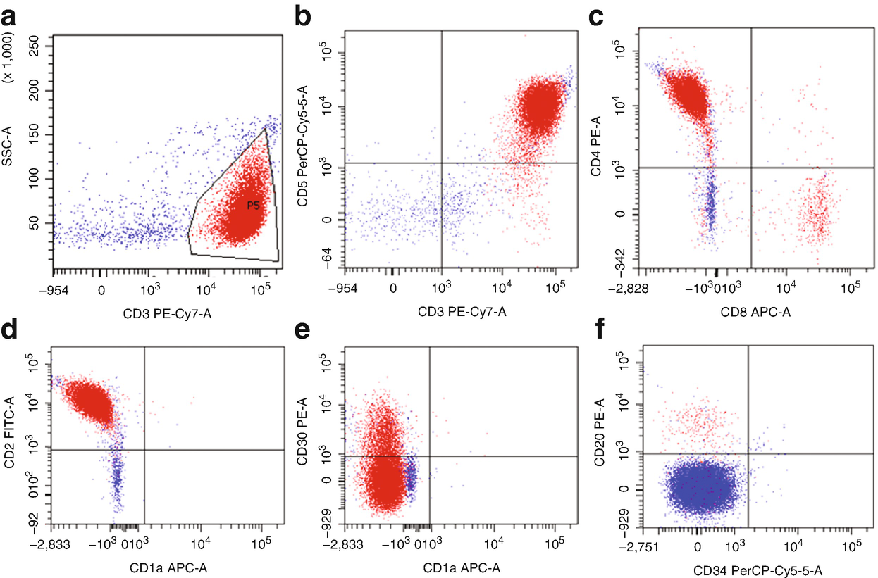
Therefore flow cytometry is an important integral part of lymphoma diagnosis even in cases where it cannot give a definitive diagnosis. The diagnosis in these cases included large cell lymphoma n3 mantle cell. After review of the clinical history and morphology a panel of markers is selected for each case by a board-certified hematopathologist.
Suspected lymphoma past history of lymphoma lymphadenopathy radiologyimaging suggests lymphoma Flow is of no value in Hodgkin lymphomas. Flow cytometry FC is usually recommended for the classification and staging of lymphomas in patients with organomegaly and atypical cells in effusions and blood after the exclusion of other possible diagnoses. Two-color analysis primarily for surface markers is currently the standard method for flow cytometry measurements in routine diagnostic studies of leukemia and lymphoma.
The concordance rate between histomorphology and flow cytometry was 915 n227. For me I can instantly tell a lymphoma from a leukaemia just by looking at the film with no flow cytometry the type conditions are very different. Learn How Mayo Clinic Can Help You.
Jevremovic D Olteanu H. Flow cytometry immunophenotyping may be useful in helping to diagnose classify treat and determine prognosis of these blood cell cancers. These can be stratified as large and small lymphocytes CD45 positive.
REQUEST FLOW CYTOMETRY ON TISSUE FOR. Discover Now Every Thing About Lymphoma. A discussion of the potential.
Three samples that came from patients who had morphologic evidence of malignant disease on biopsy two Hodgkins disease and one large cell lymphoma had flow cytometry results that were interpreted as normal. Flow Cytometry to help determine the exact type of lymphoma or exclude lymphoma This test also looks for certain molecules on the outside surface of cells by which antibodies protein molecules stick helping to identify what types of cells they are. Flow cytometric immunophenotyping is useful in diagnosing lymphoma.
FC may also have a place in the initial diagnostic investigation of aggressive lymphoma. 11 lymphs including hematogones Cytogenetics. Cytometry B Clin Cytom.
Flow cytometry is usually requested when abnormal cells are seen in the peripheral film. When using fresh tissue for flow cytometric immunophenotyping the predominant populations are lymphoid. Flow cytometry applications in the diagnosis of TNK-cell lymphoproliferative disorders.
Leukemias and lymphomas are caused by an abnormal white blood cell that begins to divide uncontrollably making numerous copies of itself clones. However flow cytometry results usually make certain lymphoma entities extremely likely and others very unlikely. This test generates a hematopathology report with a diagnosis and interpretation of findings.
This test can look at many more cells than immunohistochemistry. WBC1700uL Hb89 gdL Plt168000uL Differential count. Immunophenotyping Flow Cytometry for Hematolymphoid Neoplasia.
These cells were in the subsequent anlysis. Correlation of grade of lymphoma with flow cytometric CD19 forward scatter. Not always strictly speaking not very often.
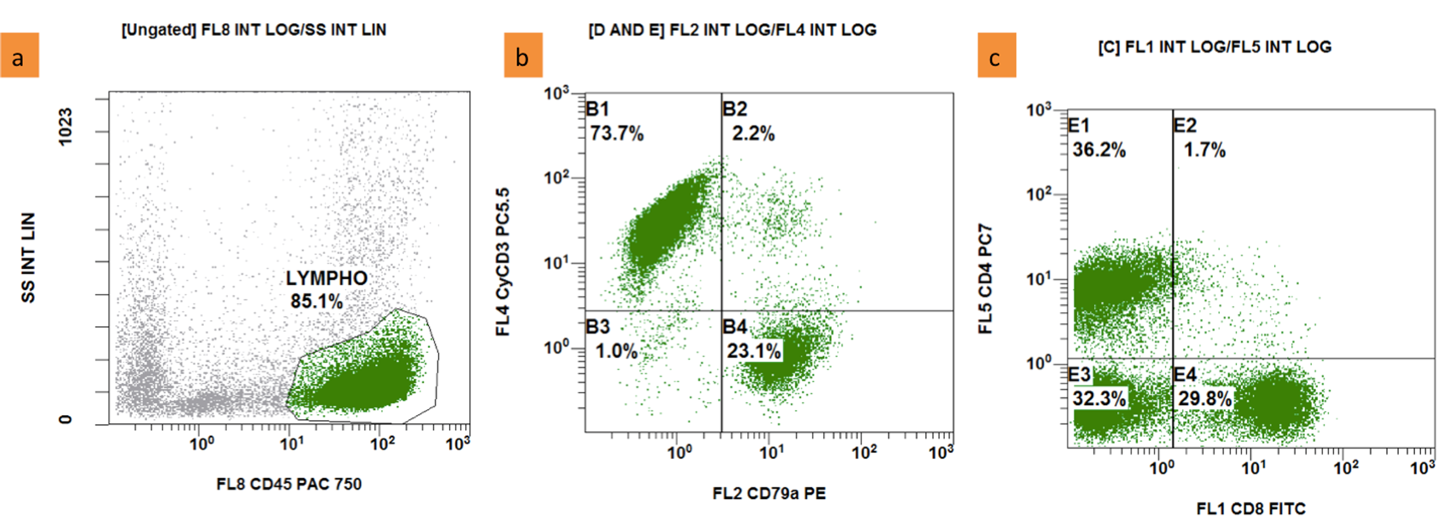
Flow cytometry FCM analysis of cerebrospinal fluid CSF is more sensitive than conventional cytology CC for diagnosis of lymphomatous meningeosis but the clinical significance of occult central nervous system CNS disease positive FCM with negative CC remains unknown. TISSUE SAMPLE REQUIREMENTS Separate fresh tissue specimen in sterile pot or in tissue preservative RPMI pink liquid preferred if sending after hours. The gating dot plot below identifies a predominant CD45 bright FS small used cells.
The advantages of flow cytometry are based largely on its ability to analyse rapidly and simultaneously multiple cell properties in a quantitative manner. The official flow cytometry labo- ratory report is most commonly an individual-lab-generated paper report form. This is especially true if initial testing showed an increased number of lymphocytes abnormal cell counts or the presence of immature blood cells.
Cureus Flow Cytometry In The Diagnosis Of Diffuse Large B Cell Lymphoma Based On Stomach Tissue Samples A Case Report

Examples Of Cd200 Expression In Mantle Cell Lymphoma By Flow Cytometry Download Scientific Diagram

Selected Flow Cytometric Immunophenotyping Plots From Fine Needle Download Scientific Diagram

Use Of Flow Cytometry In The Phenotypic Diagnosis Of Hodgkin S Lymphoma Grewal 2019 Cytometry Part B Clinical Cytometry Wiley Online Library

Flow Cytometry Results Flow Cytometric Graphs Showing Positivity For Download Scientific Diagram

Impact Of Flt3 Receptor Cd135 Detection By Flow Cytometry On Clinical Outcome Of Adult Acute Myeloid Leukemia Patients Clinical Lymphoma Myeloma And Leukemia

A D Flow Cytometry Interpretation The Neoplastic Cells Display The Download Scientific Diagram

The Current Role Of Clinical Flow Cytometry In The Evaluation Of Mature B Cell Neoplasms Seegmiller 2019 Cytometry Part B Clinical Cytometry Wiley Online Library

Use Of Flow Cytometry In The Phenotypic Diagnosis Of Hodgkin S Lymphoma Grewal 2019 Cytometry Part B Clinical Cytometry Wiley Online Library
Flow Cytometry Tutorial Flow Cytometry Data Analysis Flow Cytometry Gating Youtube
Cureus Flow Cytometry In The Diagnosis Of Diffuse Large B Cell Lymphoma Based On Stomach Tissue Samples A Case Report
Flow Cytometry Of Mature And Immature T Cell Lymphoma Springerlink

B Flow Cytometry On Peripheral Blood Revealed An Abnormal Population Download Scientific Diagram

Flow Cytometric Presentation Of A Large B Cell Lymphoma A Forward Download Scientific Diagram
International Clinical Cytometry Society

Pb Flow Cytometric Analysis Download Table

Use Of Flow Cytometry In The Phenotypic Diagnosis Of Hodgkin S Lymphoma Grewal 2019 Cytometry Part B Clinical Cytometry Wiley Online Library

Cd45 Vs Side Scatter In Clinical Sample Gating A In Clinical Flow Download Scientific Diagram

Flow Cytometric Analysis Of Representative Tissue From The Inguinal Download Scientific Diagram